Welcome to the Amira-Avizo Software Use Case Gallery
Below you will find a collection of use cases of our 3D data visualization and analysis software. These use cases include scientific publications, articles, papers, posters, presentations or even videos that show how Amira-Avizo Software is used to address various scientific and industrial research topics.
Use the Domain selector to filter by main application area, and use the Search box to enter keywords related to specific topics you are interested in.
Synergistic role of nucleotides and lipids for the self-assembly of Shs1 septin oligomers
Amira capacities for membranes and filaments segmentation in cryo-TEM images are featured on the front cover of Biochemical Journal, July 2020.
Budding yeast septins are essential for cell division and polarity. (…) [The authors] have dissected, here, for the first time, the behavior of the Shs1 protomer bound to membranes at nanometer resolution, in complex with the other septins. Using electron microscopy, [the authors] have shown that on membranes, Shs1 protomers self-assembl... Read more
Cyntia Taveneau, Rémi Blanc, Gerard Pehau-Arnaudet, Aurélie Cicco, Aurélie Bertin

Protocols for Generating Surfaces and Measuring 3D Organelle Morphology Using Amira
High-resolution 3D images of organelles are of paramount importance in cellular biology. Although light microscopy and transmission electron microscopy (TEM) have provided the standard for imaging cellular structures, they cannot provide 3D images.
However, recent technological advances such as serial block-face scanning electron microscopy (SBF-SEM) and focused ion beam scanning electron microscopy (FIB-SEM) provide the tools to create 3D images for the ultrastructural analysis of org... Read more
Edgar Garza-Lopez, Zer Vue, Prasanna Katti, Kit Neikirk, Michelle Biete, Jacob Lam, Heather K. Beasley, Andrea G. Marshall, Taylor A. Rodman, Trace A. Christensen, Jeffrey L. Salisbury, Larry Vang, Margaret Mungai, Salma Ash Shareef, Sandra A. Murray, Jianqiang Shao, Jennifer Streeter, Brian Glancy, Renata O. Pereira1, E. Dale Abel, and Antentor Hinton, Jr.
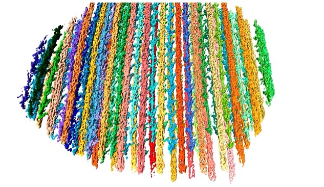
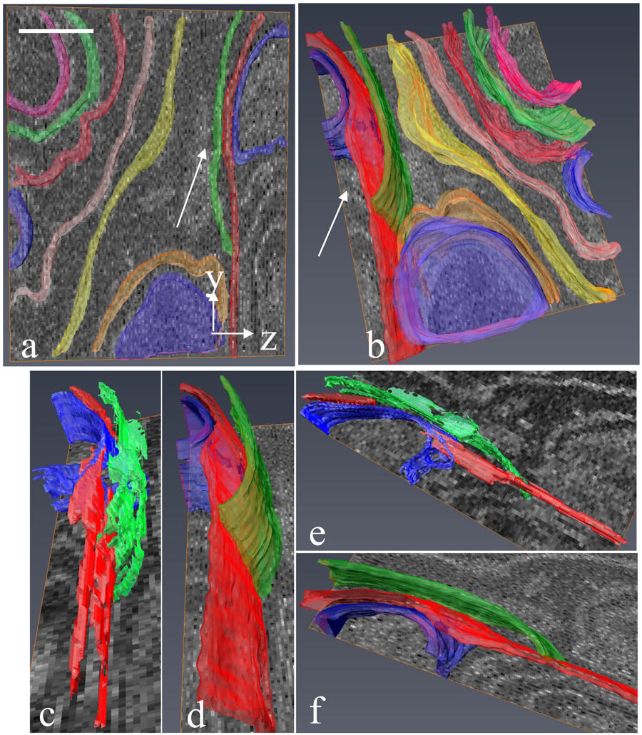
The molecular basis for sarcomere organization in vertebrate skeletal muscle
Sarcomeres are force-generating and load-bearing devices of muscles. A precise molecular picture of how sarcomeres are built underpins understanding their role in health and disease. Here, we determine the molecular architecture of native vertebrate skeletal sarcomeres by electron cryo-tomography.
Our reconstruction reveals molecular details of the three-dimensional organization and interaction of actin and myosin in the A-band, I-band, and Z-disc and demonstrates that α-actinin cros... Read more
Zhexin Wang, Michael Grange, Thorsten Wagner, Ay Lin Kho, Mathias Gautel, Stefan Raunser
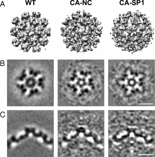
HIV-1 maturation occurs via multiple proteolytic cleavages of the Gag polyprotein, causing rearrangement of the virus particle required for infectivity. (…) How individual cleavages contribute to changes in protein structure and interactions, and how the mature, conical capsid forms, are poorly understood. Here, we employed cryoelectron tomography to determine morphology and high-resolution CA lattice structures for HIV1 derivatives in which Gag cleavage sites are mutated. These analyse... Read more
Simone Mattei, Aaron Tan, Barbel Glass, Barbara Muller, Hans-Georg Krausslich, and John A. G. Briggs
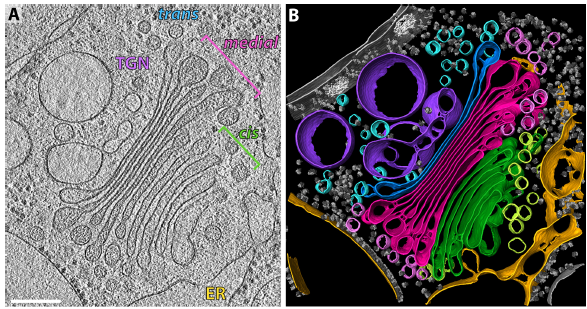
The structure of the COPI coat determined within the cell
COPI-coated vesicles mediate trafficking within the Golgi apparatus and from the Golgi to the endoplasmic reticulum. Here, we applied cryo-focused ion beam milling, cryo-electron tomography and subtomogram averaging to determine the native structure of the COPI coat within vitrified Chlamydomonas reinhardtii cells. The native algal structure resembles the in vitro mammalian structure, but additionally reveals cargo bound beneath beta’–COP. We find that all coat components disassemble... Read more
Yury S Bykov, Miroslava Schaffer, Svetlana O Dodonova, Sahradha Albert, Jurgen M Plitzko, Wolfgang Baumeister, Benjamin D Engel, John AG Briggs
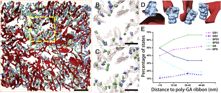
In Situ Structure of Neuronal C9orf72 Poly-GA Aggregates Reveals Proteasome Recruitment
Protein aggregation and dysfunction of the ubiquitin-proteasome system are hallmarks of many neurodegenerative diseases. Here, we address the elusive link between these phenomena by employing cryo-electron tomography to dissect the molecular architecture of protein aggregates within intact neurons at high resolution. We focus on the poly-Gly-Ala (poly-GA) aggregates resulting from aberrant translation of an expanded GGGGCC repeat in C9orf72, the most common genetic cause of amyotrophic latera... Read more
Qiang Guo, Carina Lehmer, Antonio Martinez-Sanchez, Till Rudack, Florian Beck, Hannelore Hartmann, Manuela Perez-Berlanga, Frederic Frottin, Mark S.Hipp, F. Ulrich Hartl, Dieter Edbauer, Wolfgang Baumeister, Ruben Fernandez-Busnadiego
Serial block-face electron microscopy (SBEM) provides nanoscale 3D ultrastructure of embedded and stained cells and tissues in volumes of up to 107 µm3. In SBEM, electrons with 1–3 keV energies are incident on a specimen block, from which backscattered electron (BSE) images are collected with x, y resolution of 5–10 nm in the block-face plane, and successive layers are removed by an in situ ultramicrotome. Sp... Read more
Q. He, M. Hsueh, G. Zhang, D. C. Joy & R. D. Leapman
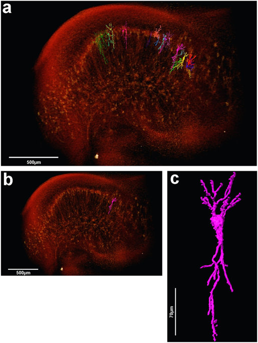
The assessment of neuronal number, spatial organization and connectivity is fundamental for a complete understanding of brain function. However, the evaluation of the three-dimensional (3D) brain cytoarchitecture at cellular resolution persists as a great challenge in the field of neuroscience. In this context, X-ray microtomography has shown to be a valuable non-destructive tool for imaging a broad range of samples, from dense materials to soft biological specimens, arisen as a new method fo... Read more
Matheus de Castro Fonseca, Bruno Henrique Silva Araujo, Carlos Sato Baraldi Dias, Nathaly Lopes Archilha, Dionísio Pedro Amorim Neto, Esper Cavalheiro, Harry Westfahl Jr, Antônio José Roque da Silva, Kleber Gomes Franchini